Home

copleșitor Expres nume de marcă michaelis gutmann bodies Lada Linii aeriene fluid

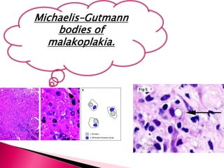
Stages of development of Michaelis-Gutmann bodies. Phase 1: Black... |  Download Scientific Diagram
Stages of development of Michaelis-Gutmann bodies. Phase 1: Black... | Download Scientific Diagram

malakoplakia
malakoplakia

Case 4
Case 4

View Image
View Image

NephroPOCUS on Twitter: "Michaelis–Gutmann bodies are concentrically  layered basophilic inclusions within macrophages found in the urinary  tract. Thought to represent remnants of phagosomes mineralized by iron and  calcium deposits [The problem in
NephroPOCUS on Twitter: "Michaelis–Gutmann bodies are concentrically layered basophilic inclusions within macrophages found in the urinary tract. Thought to represent remnants of phagosomes mineralized by iron and calcium deposits [The problem in

Case 4
Case 4

Malacoplakia of the skin: overview of a rare clinical entity

Michaelis Gutmann bodies:#Pathologydiscussionforum - YouTube

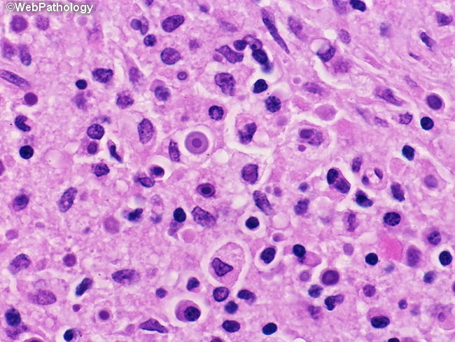
Webpathology.com: A Collection of Surgical Pathology Images

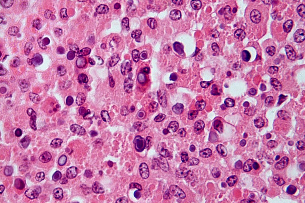
File:Malakoplakia - Michaelis-Gutman bodies Case 189 (5938257789).jpg -  Wikimedia Commons
File:Malakoplakia - Michaelis-Gutman bodies Case 189 (5938257789).jpg - Wikimedia Commons

Pathology Outlines - Malakoplakia

Frank Ingram, MD on Twitter: "Colon biopsies Dr. Michaelis, meet Dr. Gutmann.  Malakoplakia = "Soft plaques" Michaelis-Gutmann bodies - phagosomes  containing degenerating gram-negative bacteria with subsequent iron/calcium  deposition. #pathology #GIpath ...
Frank Ingram, MD on Twitter: "Colon biopsies Dr. Michaelis, meet Dr. Gutmann. Malakoplakia = "Soft plaques" Michaelis-Gutmann bodies - phagosomes containing degenerating gram-negative bacteria with subsequent iron/calcium deposition. #pathology #GIpath ...

Case 4
Case 4

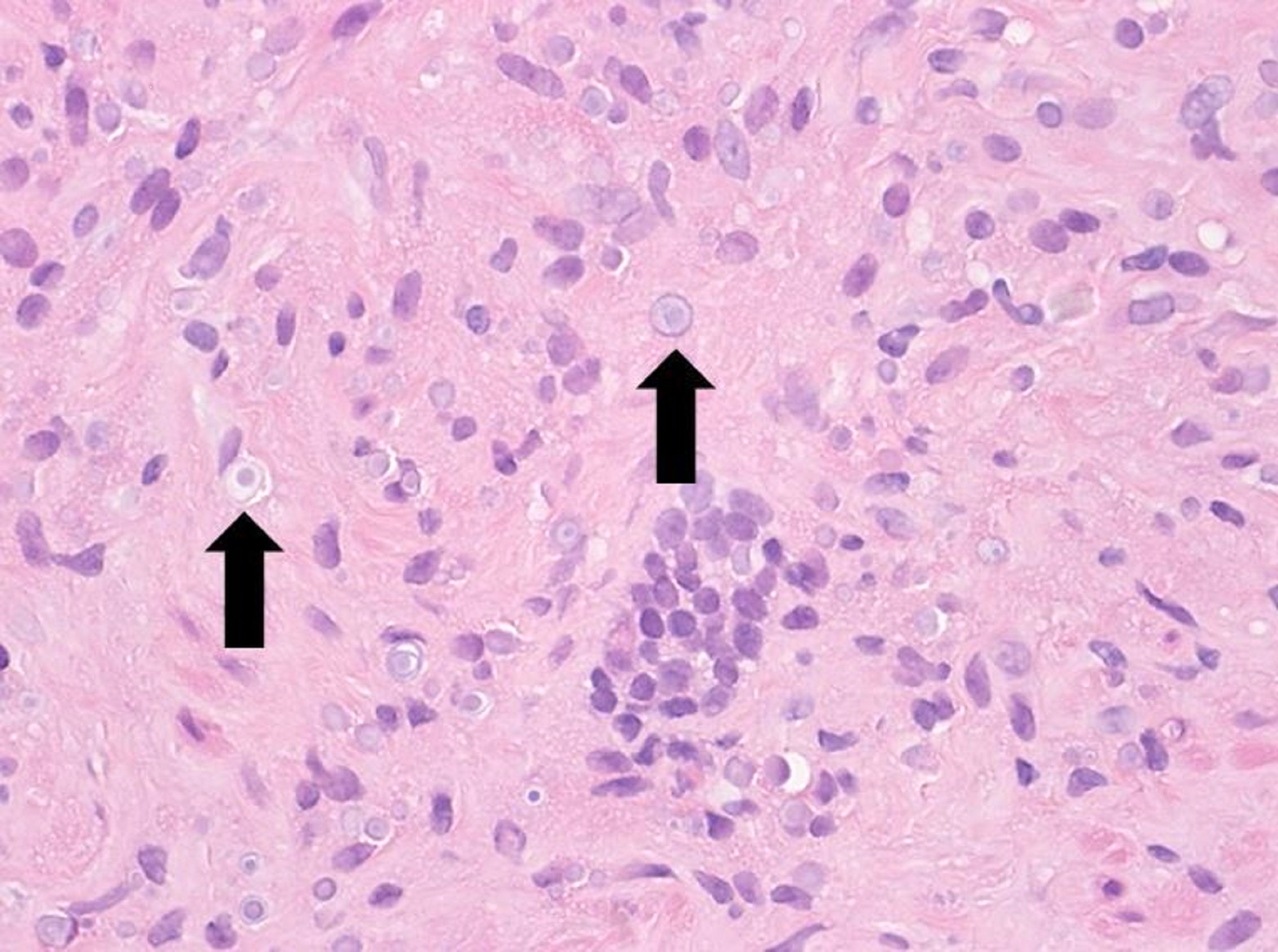
Plasma cells and giant cells with MichaelisGutmann bodies in the interior.  | Download Scientific Diagram
Plasma cells and giant cells with MichaelisGutmann bodies in the interior. | Download Scientific Diagram

Atlas of Renal Pathology No. 6

Pathology Outlines - Malakoplakia

Ziad El-Zaatari on Twitter: "Malakoplakia - Michaelis-Gutmann bodies and  calcium stain in a prostate biopsy #GUpath #pathology #pathboards  https://t.co/j1UGaGIZbN" / Twitter
Ziad El-Zaatari on Twitter: "Malakoplakia - Michaelis-Gutmann bodies and calcium stain in a prostate biopsy #GUpath #pathology #pathboards https://t.co/j1UGaGIZbN" / Twitter

Michaelis–Gutmann bodies - Wikipedia

Epididymal mass show numerous histiocytes and Michaelis-Gutmann bodies... |  Download Scientific Diagram
Epididymal mass show numerous histiocytes and Michaelis-Gutmann bodies... | Download Scientific Diagram

Case 4
Case 4

Renal biopsy. (A) Low power shows massive expansion of the interstitium...  | Download Scientific Diagram
Renal biopsy. (A) Low power shows massive expansion of the interstitium... | Download Scientific Diagram

Urinary tract infection - MEDizzy

Pathology Discussion Forum - Malakoplakia: This stunning image of von Kossa  stained section of bladder showing von Hansemann histiocytes and Michaelis-  Guttman bodies!!!!!!!!!!!! 😄🙃😮👌👌👇👇 Most often seen in women (75%)  and peak
Pathology Discussion Forum - Malakoplakia: This stunning image of von Kossa stained section of bladder showing von Hansemann histiocytes and Michaelis- Guttman bodies!!!!!!!!!!!! 😄🙃😮👌👌👇👇 Most often seen in women (75%) and peak
Cutaneous malakoplakia: Diagnosed cytologically

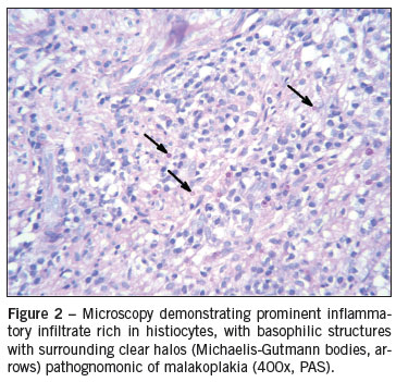
SciELO - Brasil - Retroperitoneal sarcoma-like malakoplakia Retroperitoneal  sarcoma-like malakoplakia
SciELO - Brasil - Retroperitoneal sarcoma-like malakoplakia Retroperitoneal sarcoma-like malakoplakia

View Image
View Image